Neuron-glial antigen2 antibody - 481 002

The NG2 is a valuable marker for several incompletely differentiated precursor cells
Rabbit polyclonal antiserum
Cat. No.: 481 002
Amount: 200 µl
Price: $360.00
Cat. No. 481 002 200 µl antiserum, lyophilized. For reconstitution add 200 µl H2O, then aliquot and store at -20°C until use.
Antibodies should be stored at +4°C when still lyophilized. Do not freeze!
Applications
 
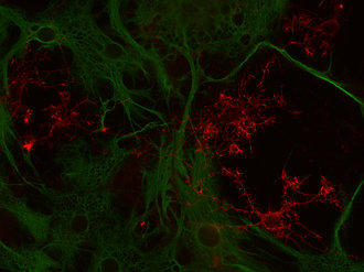
WB: 1 : 500 up to 1 : 1000 (AP-staining) gallery  
IP: yes
ICC: 1 : 500 (see remarks) gallery  
IHC: not recommended
IHC-P: not recommended
IHC-Fr: not recommended

Western blot (WB); separation of proteins by PAGE and subsequent transfer to a membrane. Detection of target molecules is carried out with antibodies. Some antibodies require special sample preparation steps. For details, please refer to the “Remarks” section.

Immunoprecipitation (IP); Immunoisolation or pulldown of a target molecule using an antibody. For details and product specific hints, please refer to the ”Remarks” section.

Immunocytochemistry (ICC) on 4% PFA fixed cells. Immunoreactivity is usually revealed by fluorescence. Some antibodies require special fixation methods. For details, please refer to the “Remarks” section.

Immunohistochemistry (IHC) on 4% PFA perfusion fixed tissue with 24h PFA post fixation. Immunoreactivity is usually revealed by fluorescence or a chromogenic substrate. Some antibodies require special fixation methods or antigen retrieval steps. For details, please refer to the ”Remarks” section.

Immunohistochemistry (IHC-P) of formalin fixed, paraffin embedded (FFPE) tissue (some antibodies require special antigen retrieval steps, please refer to the ”Remarks” section). Immunoreactivity is usually revealed by fluorescence or a chromogenic substrate.

Immunohistochemistry on fresh frozen (IHC-Fr) cryo-tissue-sections. In contrast to standard PFA perfusion fixed tissues, fresh frozen cryo-tissue-sections can be variably postfixed with alcohols, acetone or PFA. Alcohol or acetone fixation is e.g. of advantage for antigens masked by PFA crosslinking. For recommended postfixation, please refer to the ”Remarks” section. Immunoreactivity is usually revealed by fluorescence or a chromogenic substrate.

Immunogen Synthetic peptide mix corresponding to residues near the carboxy and amino terminus of mouse Neuron-glial antigen 2 (UniProt Id: Q8VHY0)
Reactivity Reacts with: mouse (Q8VHY0), rat (Q00657).
Other species not tested yet.
Remarks

ICC: Works on PFA and methanol fixed cells.

This antibody shows weak unspecific nuclear staining of neurons.

Data sheet 481_002.pdf
Cat. No.: 481 002
Amount: 200 µl
Price: $360.00
Background

The NG2 proteoglycan is a type I membrane protein that is expressed by a variety of immature cells of several embryonic tissue origins including glia, muscle progenitor cells, and pericytes (1). In the central nervous system, expression of NG2 was originally thought to specify oligodendroglial progenitor cells, but more recent data suggest that NG2-expressing cells encompass a wider range of immature glial cells in white and gray matter. These include glia that make synaptic-like contacts with neurons in the hippocampus and cerebellum (2) and glial cells specifically associated with the nodes of Ranvier (3). Interestingly, many NG2-positive cells are both proliferative and motile or exhibit local process motility (4, 5).