| Cat. No. HS-384 008 |
100 µl purified recombinant IgG, lyophilized. Albumin and azide were added for stabilization. For reconstitution add 100 µl H2O. Then aliquot and store at -20°C to -80°C until use. Antibodies should be stored at +4°C when still lyophilized. Do not freeze! |
| Applications | |
| Clone | Rb298G2 |
| Subtype | IgG1 (κ light chain) |
| Immunogen | synthetic peptide corresponding to residues surrounding AA 1000 of mouse CD11b (UniProt Id: P05555) |
| Reactivity |
Reacts with: mouse (P05555). No signal: human (P11215), rat (G3V8L7). Other species not tested yet. |
| Remarks |
This antibody is a chimeric antibody based on the monoclonal rat antibody clone 298G2H5. The constant regions of the heavy and light chains have been replaced by rabbit specific sequences. Therefore, the antibody can be used with standard anti-rabbit secondary reagents. The antibody has been expressed in mammalian cells. |
| Data sheet | hs-384_008.pdf |

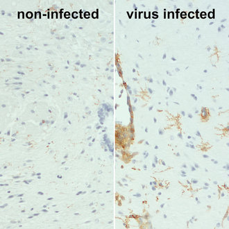
Increased CD11b expression represents microglial activation in virus infected mouse brain
CD11b also called integrin alpha-M (ITGAM) is one protein subunit that forms together with CD18 the heterodimeric integrin αMβ2 complex, also known as macrophage-1 antigen (Mac-1) or complement receptor 3 (CR3) (1). αMβ2 is expressed on polymorphonuclear neutrophils (PMN), monocytes, macrophages, some subsets of cytotoxic T lymphocytes, and NK cells. Antibodies against CD11b are frequently used to identify macrophages and microglia, however not all tissue-resident macrophages are CD11b positive. In the murine liver CD11b expression is rare and almost exclusively found on F4/80 negative cells (2). In the murine spleen CD11b+ cells are less numerous than F4/80+ cells, co-expression of CD11b and F4/80 is also rare and CD11b+ cells tend to be closer to the marginal zone (2). CD11b upregulation on residential alveolar macrophages is a marker of acute and chronic lung inflammation in mice (3). Also, in the brain CD11b is markedly increased during microglial activation (4).