| Cat. No. HS-384 017 |
200 µl purified IgG, lyophilized. Albumin and azide were added for stabilization. For reconstitution add 200 µl H2O. Then aliquot and store at -20°C to -80°C until use. Antibodies should be stored at +4°C when still lyophilized. Do not freeze! |
| Applications | |
| Clone | 194B8D5 |
| Subtype | IgG2b (κ light chain) |
| Immunogen | Synthetic peptide corresponding to residues near the carboxy terminus of human CD11b (UniProt Id: P11215) |
| Reactivity |
Reacts with: human (P11215). No signal: mouse (P05555). Other species not tested yet. |
| Remarks |
IHC-P: This antibody shows weak non-specific cross-reactivity to murine macrophages after antigen retrieval at very high temperatures (121°, pressure cooker). Use appropriate controls when optimizing your IHC-P protocol for detection of human cells in humanized mouse models. |
| Data sheet | hs-384_017.pdf |

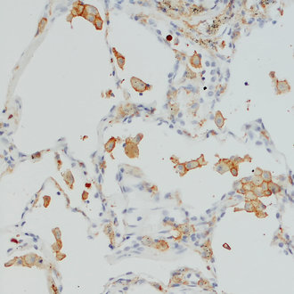
Indirect immunostaining of human lung section with rat anti-CD11b detects low CD11b expression in alveolar macrophages.
CD11b also called integrin alpha-M (ITGAM) is one protein subunit that forms together with CD18 the heterodimeric integrin αMβ2 complex, also known as macrophage-1 antigen (Mac-1) or complement receptor 3 (CR3) (1). αMβ2 is expressed on polymorphonuclear neutrophils (PMN), monocytes, macrophages, some subsets of cytotoxic T lymphocytes, and NK cells. Antibodies against CD11b are frequently used to identify macrophages and microglia, however not all tissue-resident macrophages are CD11b positive. In the murine liver CD11b expression is rare and almost exclusively found on F4/80 negative cells (2). In the murine spleen CD11b+ cells are less numerous than F4/80+ cells, co-expression of CD11b and F4/80 is also rare and CD11b+ cells tend to be closer to the marginal zone (2). CD11b upregulation on residential alveolar macrophages is a marker of acute and chronic lung inflammation in mice (3). Also, in the brain CD11b is markedly increased during microglial activation (4).