F4/80 antibody - HS-397 017

F4/80 is a marker protein for murine macrophages
Rat monoclonal purified IgG
Cat. No.: HS-397 017
Amount: 200 µl
Price: $420.00
Cat. No. HS-397 017 200 µl purified IgG, lyophilized. Albumin and azide were added for stabilization. For reconstitution add 200 µl H2O. Then aliquot and store at -20°C to -80°C until use.
Antibodies should be stored at +4°C when still lyophilized. Do not freeze!
Applications
 
WB: 1 : 250 (AP staining) gallery  
IP: not tested yet
ICC: not tested yet
IHC: 1 : 500 gallery  
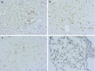
IHC-P: 1 : 100 gallery  
IHC-Fr: 1 : 500 (see remarks) gallery  

Western blot (WB); separation of proteins by PAGE and subsequent transfer to a membrane. Detection of target molecules is carried out with antibodies. Some antibodies require special sample preparation steps. For details, please refer to the “Remarks” section.

Immunoprecipitation (IP); Immunoisolation or pulldown of a target molecule using an antibody. For details and product specific hints, please refer to the ”Remarks” section.

Immunocytochemistry (ICC) on 4% PFA fixed cells. Immunoreactivity is usually revealed by fluorescence. Some antibodies require special fixation methods. For details, please refer to the “Remarks” section.

Immunohistochemistry (IHC) on 4% PFA perfusion fixed tissue with 24h PFA post fixation. Immunoreactivity is usually revealed by fluorescence or a chromogenic substrate. Some antibodies require special fixation methods or antigen retrieval steps. For details, please refer to the ”Remarks” section.

Immunohistochemistry (IHC-P) of formalin fixed, paraffin embedded (FFPE) tissue (some antibodies require special antigen retrieval steps, please refer to the ”Remarks” section). Immunoreactivity is usually revealed by fluorescence or a chromogenic substrate.

Immunohistochemistry on fresh frozen (IHC-Fr) cryo-tissue-sections. In contrast to standard PFA perfusion fixed tissues, fresh frozen cryo-tissue-sections can be variably postfixed with alcohols, acetone or PFA. Alcohol or acetone fixation is e.g. of advantage for antigens masked by PFA crosslinking. For recommended postfixation, please refer to the ”Remarks” section. Immunoreactivity is usually revealed by fluorescence or a chromogenic substrate.

Clone 167B3
Subtype IgG2a (κ light chain)
Immunogen Synthetic peptide corresponding to AA 28 to 42 from mouse F4/80 (UniProt Id: Q61549)
Reactivity Reacts with: mouse (Q61549).
No signal: rat.
Other species not tested yet.
Remarks

IHC-Fr: PFA and MeOH fixation are recommended.

Data sheet hs-397_017.pdf
Cat. No.: HS-397 017
Amount: 200 µl
Price: $420.00

F4/80 positive cells in murine FFPE tissues

Background
The murine macrophage cell surface glycoprotein F4/80 is a member of the epidermal growth factor-seven transmembrane (EGF-TM7) family. It is involved in the generation of antigen-specific efferent regulatory T cells that suppress antigen-specific immunity but not required for the development and distribution of tissue macrophages.
Although F4/80 is widely used as a marker of murine macrophage populations it is not equivalently expressed across tissue-specific macrophage lineages: e.g. red pulp macrophages of the spleen and Kupffer cells of the liver are F4/80-positive, white pulp and marginal zone macrophages of the spleen are F4/80-negative and alveolar macrophages are F4/80dim.
F4/80 expression is not restricted to macrophages, but also found in murine Epidermal Langerhans Cells; dendritic cells of the skin. The human ortholog of F4/80, EGF-like module containing mucin-like hormone receptor (EMR)1, is absent on mononuclear phagocytic cells including monocytes, macrophages, and myeloid dendritic cells and seems to be highly specific for eosinophils in humans.