Cat. No.: HS-413 008
Amount: 100 µl
Price:
$420.00
| Cat. No. HS-413 008 |
100 µl purified recombinant IgG, lyophilized. Albumin and azide were added for stabilization. For reconstitution add 100 µl H2O. Then aliquot and store at -20°C to -80°C until use. Antibodies should be stored at +4°C when still lyophilized. Do not freeze! |
| Applications | |
| Clone | Rb281A6 |
| Subtype | IgG1 (κ light chain) |
| Immunogen | Synthetic peptide corresponding to residues surrounding AA 55 of human CD3e (UniProt Id: P07766) |
| Reactivity |
Reacts with: human (P07766). No signal: mouse (P22646). Other species not tested yet. |
| Remarks |
This antibody is a chimeric antibody based on the monoclonal rat antibody clone 281A6F1. The constant regions of the heavy and light chains have been replaced by rabbit specific sequences Therefore, the antibody can be used with standard anti-rabbit secondary reagents. The antibody has been expressed in mammalian cells. |
| Data sheet | Datasheet hs-413_008 |
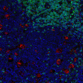
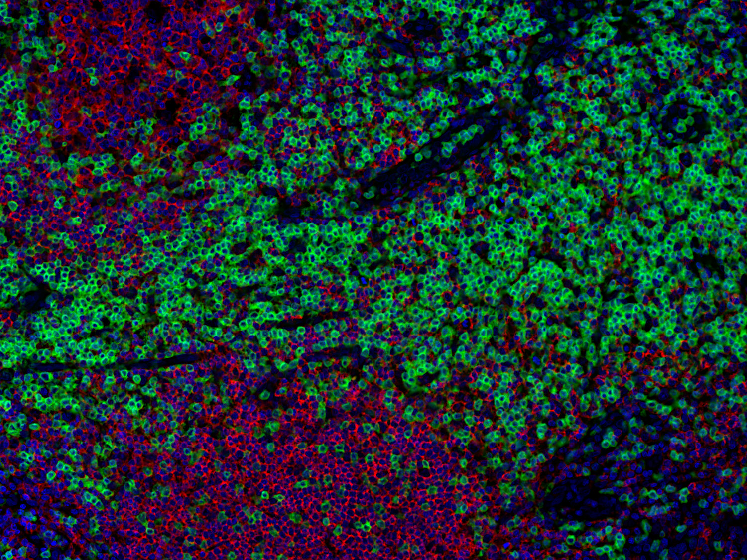
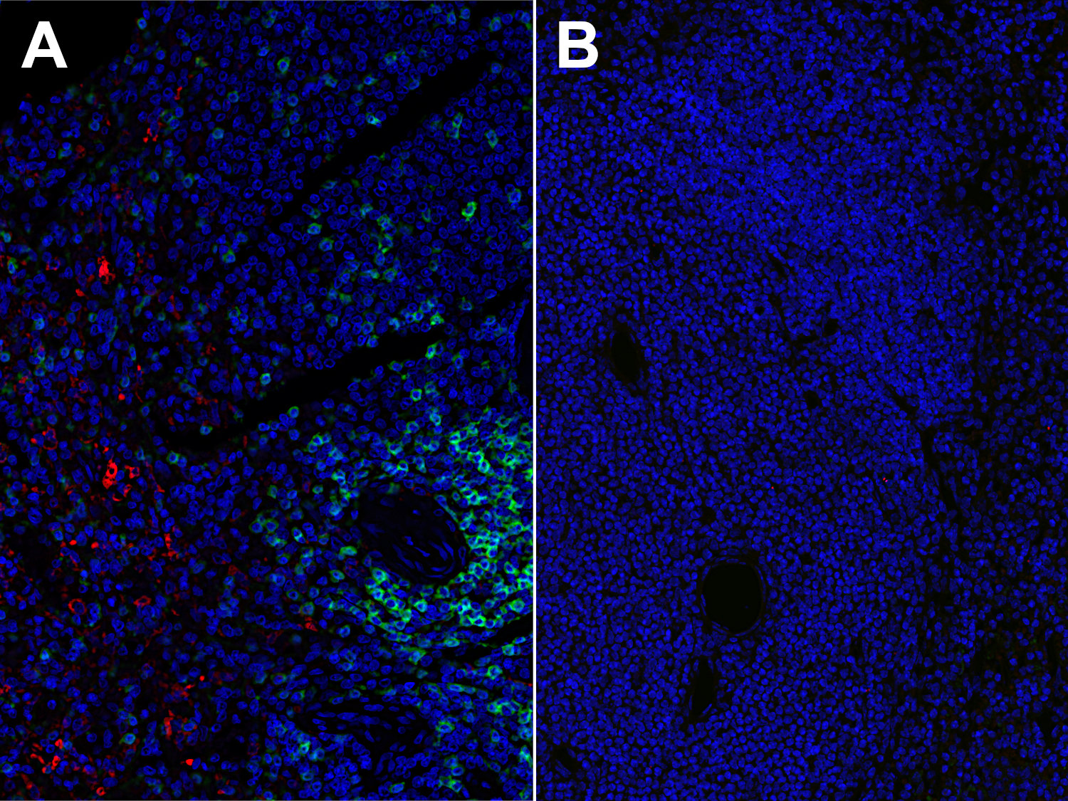
Immunofluorescence image of a human FFPE tonsil section stained for CD3e (T cells, green) and CD68 (macrophages, red)

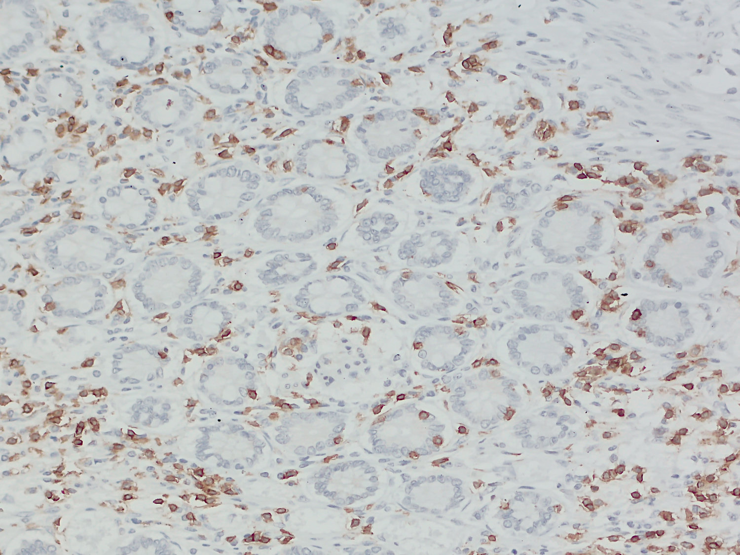
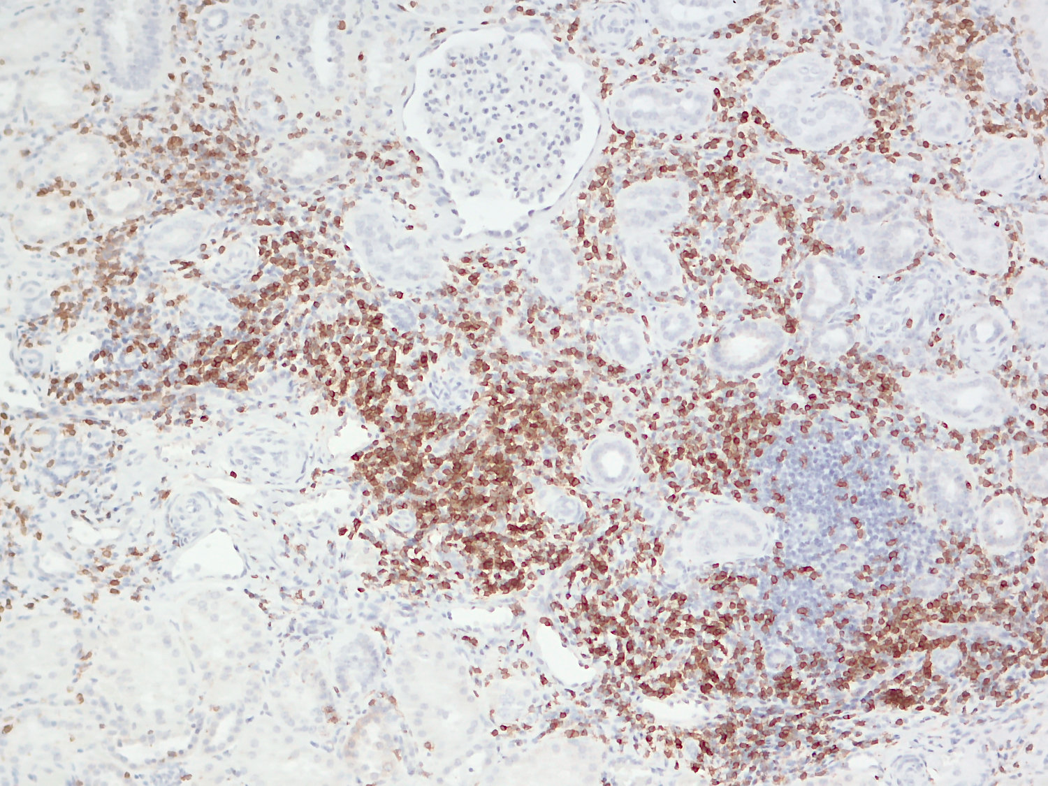
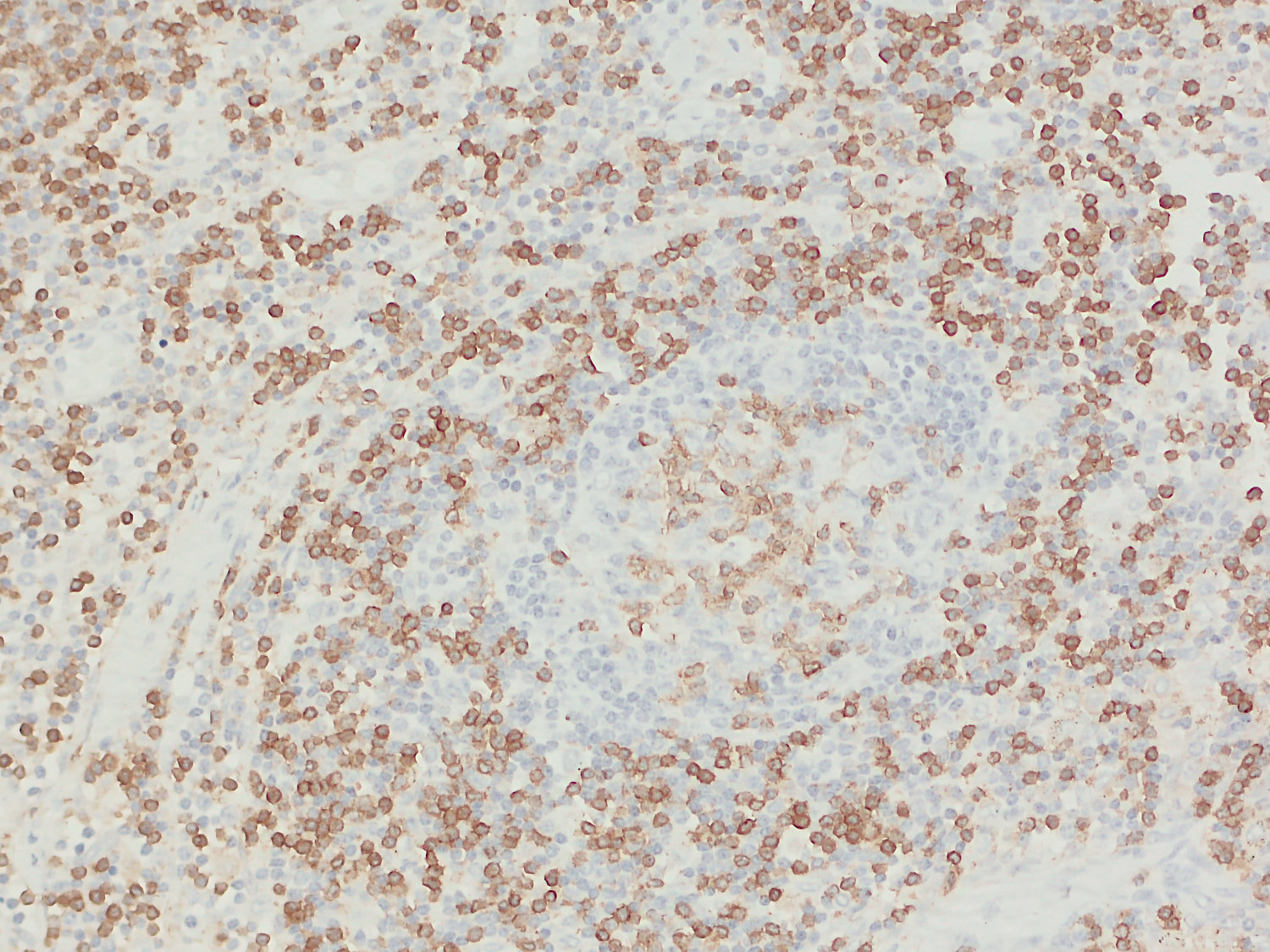
Cluster of differentiation 3 (CD3) is a defining feature of cells belonging to the T cell lineage. It is composed of the four subunits CD3 gamma, CD3 delta, CD3 epsilon (CD3e) and CD3 zeta, that form a multimeric protein complex. This complex associates with the T cell receptor (TCR) and serves as a T cell co-receptor. The CD3 molecules contain immunoreceptor tyrosine-based activation motifs (ITAMs) that serve as the nucleating point for the intracellular signal transduction machinery upon TCR engagement. TCR/CD3 signaling is central to the initiation of antigen-specific T cell responses to pathogens and vaccines, as well as transplanted tissues, tumors, and autoantigens. CD3 is initially expressed in the cytoplasm of pro-thymocytes. During T cell maturation the expression of CD3 migrates to the cell-membrane. The specific appearance at all stages of T cell development make CD3 a useful immunohistochemical marker for T cells in tissue sections. In the clinical setting, CD3 is a relevant marker for the classification of malignant lymphomas and leukemias as the antigen remains present in almost all T-cell lymphomas and leukemias. It can also be used to detect T cells in celiac disease, lymphocytic and collagenous colitis.
Certificates
ISO 9001 2015 Quality Management System and Green Lab Platinum certification level for sustaining laboratory processes.

Newsletter
Sign up for our newsletter and get the latest updates and news.