| Cat. No. HS-413 103 |
200 µl specific antibody, lyophilized. Affinity purified with the immunogen. Albumin and azide were added for stabilization. For reconstitution add 200 µl H2O. Then aliquot and store at -20°C to -80°C until use. Antibodies should be stored at +4°C when still lyophilized. Do not freeze! |
| Applications | |
| Immunogen | Synthetic peptide corresponding to AA 44 to 64 from mouse CD3e (UniProt Id: P22646) |
| Reactivity |
Reacts with: mouse (P22646). No signal: human (P07766), rat (D4A5M2). Other species not tested yet. |
| Remarks |
IHC: Antigen retrieval with citrate buffer pH 6 is required. |
| Data sheet | hs-413_103.pdf |

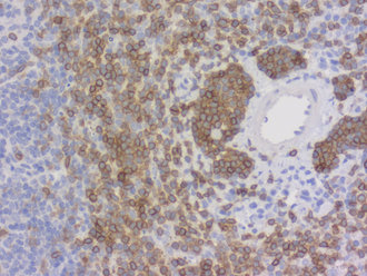
Detection of CD3e positive cells in FFPE mouse spleen section
Cluster of differentiation 3 (CD3) is a defining feature of cells belonging to the T cell lineage. It is composed of the four subunits CD3 gamma, CD3 delta, CD3 epsilon (CD3e) and CD3 zeta, that form a multimeric protein complex. This complex associates with the T cell receptor (TCR) and serves as a T cell co-receptor. The CD3 molecules contain immunoreceptor tyrosine-based activation motifs (ITAMs) that serve as the nucleating point for the intracellular signal transduction machinery upon TCR engagement. TCR/CD3 signaling is central to the initiation of antigen-specific T cell responses to pathogens and vaccines, as well as transplanted tissues, tumors, and autoantigens. CD3 is initially expressed in the cytoplasm of pro-thymocytes. During T cell maturation the expression of CD3 migrates to the cell-membrane. The specific appearance at all stages of T cell development make CD3 a useful immunohistochemical marker for T cells in tissue sections. In the clinical setting, CD3 is a relevant marker for the classification of malignant lymphomas and leukemias as the antigen remains present in almost all T-cell lymphomas and leukemias. It can also be used to detect T cells in celiac disease, lymphocytic and collagenous colitis.