| Cat. No. HS-439 017 |
100 µg purified IgG, lyophilized. Albumin and azide were added for stabilization. For reconstitution add 100 µl H2O to get a 1mg/ml solution in PBS. Then aliquot and store at -20°C to -80°C until use. Antibodies should be stored at +4°C when still lyophilized. Do not freeze! |
| Applications | |
| Clone | 26D7E5 |
| Subtype | IgG2b (κ light chain) |
| Immunogen | Synthetic peptide corresponding to residues near the carboxy terminus of mouse CD19 (UniProt Id: P25918) |
| Reactivity |
Reacts with: mouse (P25918). No signal: human (P15391), rat. Other species not tested yet. |
| Remarks |
IHC-Fr: The following fixatives are possible: methanol, 4% formaldehyde/PFA |
| Data sheet | hs-439_017.pdf |

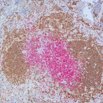
Chromogenic double staining for CD19 (DAB) and CD3e (RED) visualized T-cell and B-cell populations in the mouse spleen
CD19 (Cluster of Differentiation 19) is a B cell-restricted signal-transduction molecule that plays an important role in the regulation of development, activation, and differentiation of B-lymphocytes. CD19 is considered as a biomarker for B-cells because of its continued expression from very early B cell development stages, being evident already on pro-B cells and on all later B cell stages, until plasma cell terminal differentiation, when its expression is lost. In complex with CD21 (complement receptor-2), CD81 and CD225 (Leu-13), CD19 functions as a dominant signaling receptor on the surface of mature B cells (1). CD19 is instrumental in B cell homeostasis and lowers the threshold of B cell receptor crosslinking necessary to effect B-cell activation and sustain proliferation upon antigen encounter (2). Dysregulated CD19 expression has been implicated in several autoimmune diseases and CD19 is expressed in most acute lymphoblastic leukemias (ALL), chronic lymphocytic leukemias (CLL) and other B cell lymphomas (3). Therefore, CD19 has gained attention as a potential target in the therapy of B-cell malignancies (4).