| Cat. No. HS-460 008 |
100 µl purified recombinant IgG, lyophilized. Albumin and azide were added for stabilization. For reconstitution add 100 µl H2O. Then aliquot and store at -20°C to -80°C until use. Antibodies should be stored at +4°C when still lyophilized. Do not freeze! |
| Applications | |
| Clone | Rb186F9 |
| Subtype | IgG1 (κ light chain) |
| Immunogen | synthetic peptide corresponding to residues near the carboxy terminus of human CD68 (UniProt Id: P34810) |
| Reactivity |
Reacts with: human (P34810). No signal: mouse (P31996). Other species not tested yet. |
| Remarks |
This antibody is a chimeric antibody based on the monoclonal rat antibody clone 186F9B4. The constant regions of the heavy and light chains have been replaced by rabbit specific sequences. Therefore, the antibody can be used with standard anti-rabbit secondary reagents. The antibody has been expressed in mammalian cells. |
| Data sheet | hs-460_008.pdf |

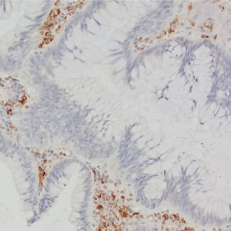
CD68 staining detects macrophages in human colon adenoma
CD68, also called Lamp-4 in humans or macrosialin in mice, is a highly glycosylated type I transmembrane protein that belongs to the lysosome associated membrane protein (LAMP) family (1). CD68 expression is increased in cells associated with elevated phagocytic and degradative activity. High CD68 expression is detected in cells of the mononuclear phagocyte lineage including macrophages, osteoclasts, and myeloid dendritic cells (2). CD68 is a marker of activated microglia and only expressed at low levels in resting microglia (3). Staining for CD68 is predominantly intracellular, only 10 -15% of it is found on the cell surface. In oncology research, CD68 is the major biomarker for quantification of tumor-associated macrophages (TAMs) (4). High infiltration of CD68+ macrophages is an independent prognostic factor for overall survival in several tumor entities (5).