| Cat. No. HS-467 003 |
50 µg specific antibody, lyophilized. Affinity purified with the immunogen. Albumin and azide were added for stabilization. For reconstitution add 50 µl H2O to get a 1mg/ml solution in PBS. Then aliquot and store at -20°C to -80°C until use. Antibodies should be stored at +4°C when still lyophilized. Do not freeze! |
| Applications | |
| Reactivity |
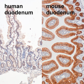
Reacts with: mouse (P09803), rat. No signal: human. Other species not tested yet. |
| Remarks |
IHC: Antigen retrieval with citrate buffer pH 6 is required. |
| Data sheet | hs-467_003.pdf |

Indirect immunostaining of human and mouse duodenum sections using mouse-E-cadherin specific antibody.
Epithelial cadherin (E-cadherin) also known as Cadherin-1, CAM 120/80 or uvomorulin belongs together with neuronal (N) cadherin to the type I classical cadherins, transmembrane proteins that function in calcium-dependent cell-cell adhesion (1). In normal tissues, E-cadherin is expressed by most epithelial cells. A distinct distribution of E-cadherin expression is found in the kidney, where only distal tubuli show E-cadherin expression and in the placenta, where only the cytotrophoblastic layer stains positive for E-cadherin (2). In the human normal adult nervous system E-cadherin expression is limited to the arachnoid membrane, whereas in mice E-cadherin is also expressed in neural stem cells, where E-cadherin regulates self-renewal (3). E-cadherin is a potent tumor suppressor and the so-called “cadherin switch” - downregulation of E-cadherin while N-cadherin is upregulated - is often found in malignant epithelial cancers. This Epithelial-to-Mesenchymal Transition (EMT) has been shown to be crucial in tumorigenesis where the EMT program enhances metastasis, chemoresistance and tumor stemness (4). However, also E-cadherin upregulation in malignancies derived from E-cadherin negative normal tissues tend to be linked to unfavorable tumor phenotype and disease outcome (2). In syngeneic mouse tumors E-cadherin expression is typically lower than in human tumors suggesting that syngeneic mouse models have a more mesenchymal-like tumor cellular phenotype (5).