Cat. No.: HS-477 103
Amount: 50 µg
Price:
$355.00
| Cat. No. HS-477 103 |
50 µg specific antibody, lyophilized. Affinity purified with the immunogen. Albumin and azide were added for stabilization. For reconstitution add 50 µl H2O to get a 1mg/ml solution in PBS. Then aliquot and store at -20°C to -80°C until use. Antibodies should be stored at +4°C when still lyophilized. Do not freeze! |
| Applications | |
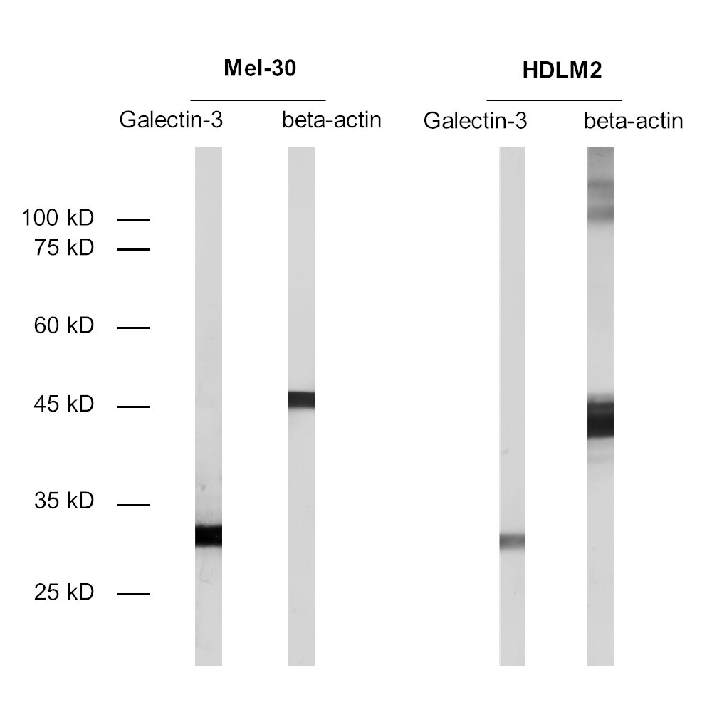
| Immunogen | Synthetic peptide corresponding to residues near the amino terminus of human Galectin-3 (UniProt Id: P17931) |
| Reactivity |
Reacts with: human (P17931). No signal: mouse (P16110). Other species not tested yet. Predicted to cross-react with monkey due to high sequence homology. |
| Remarks |
ICC: 4% formaldehyde/PFA fixation is recommended. |
| Data sheet | Datasheet hs-477_103 |
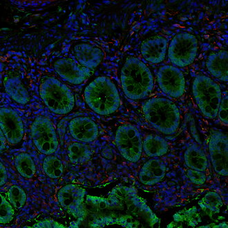
Fluorescent double staining for Galectin-3 (green) and IBA1 (red) in a human small intestine
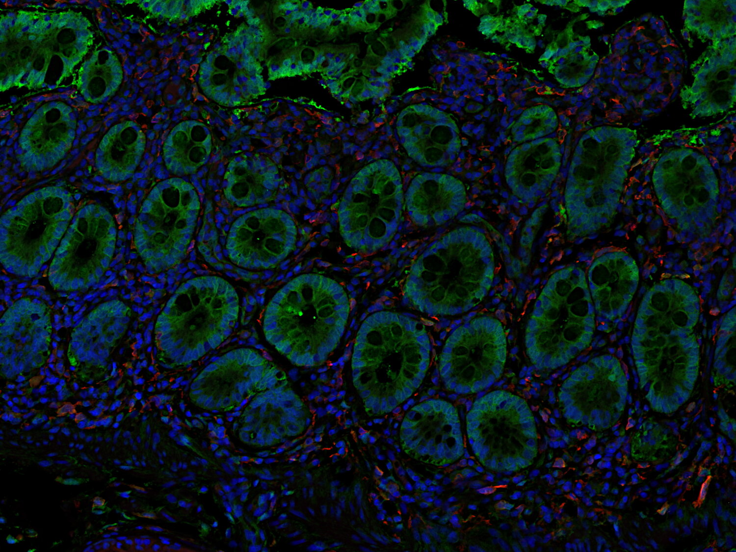
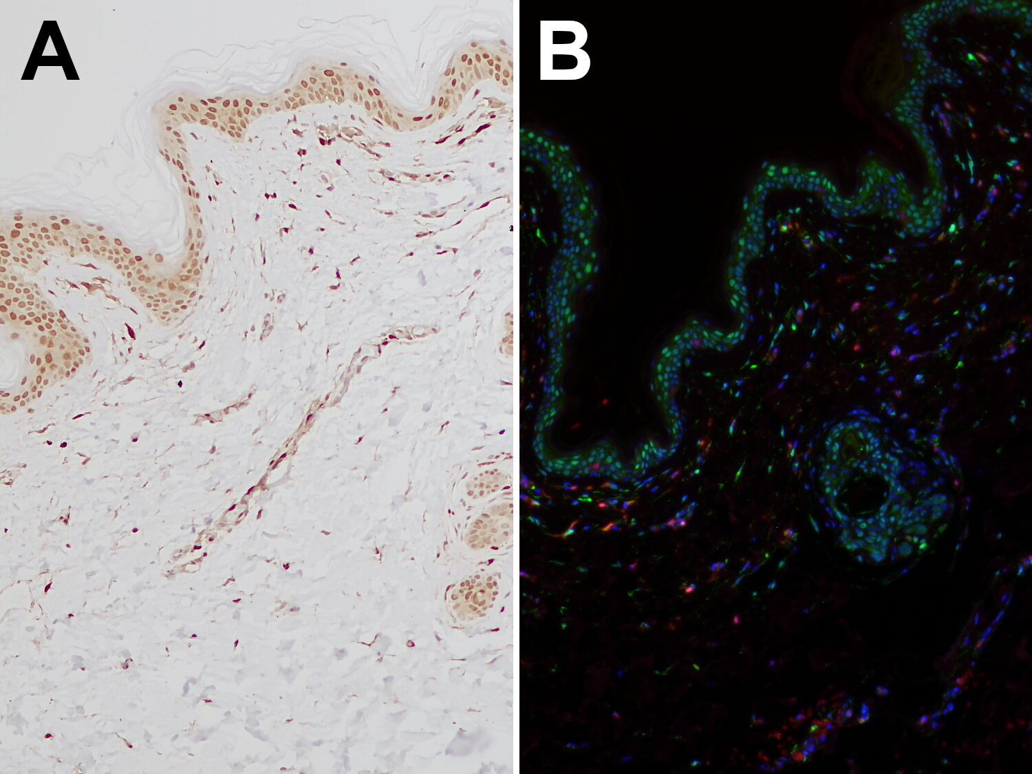
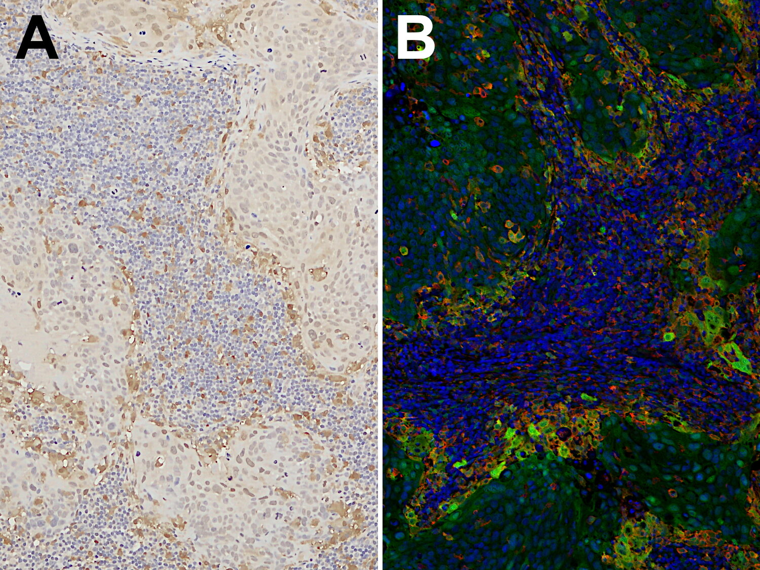
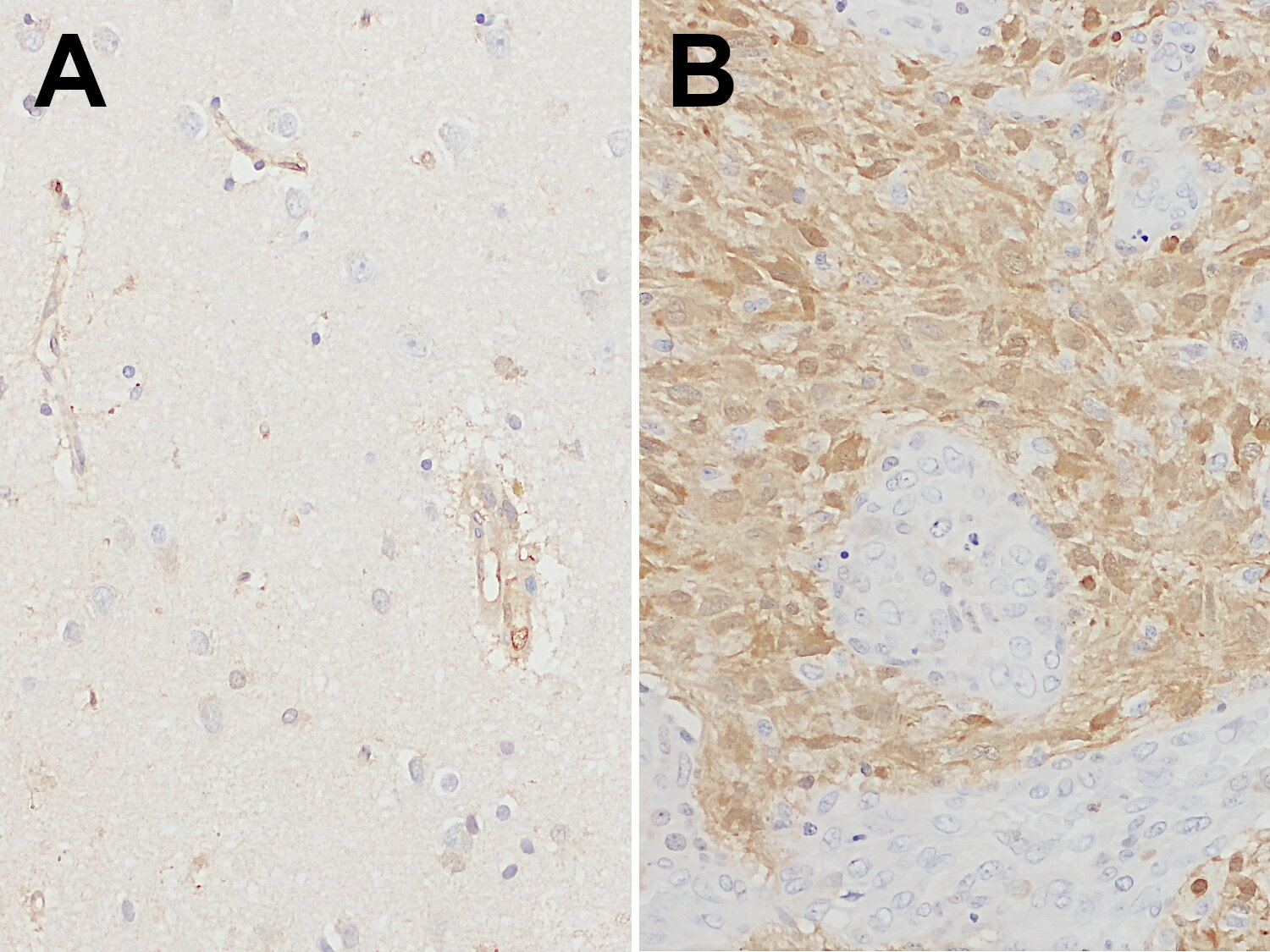
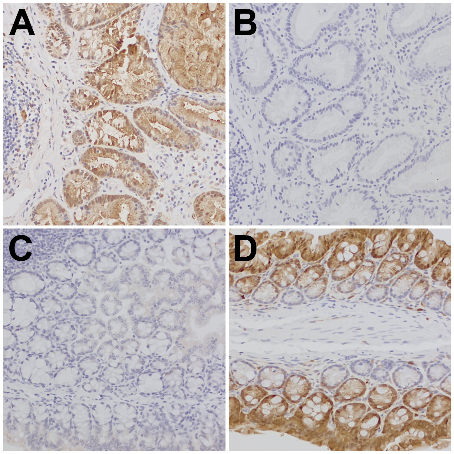
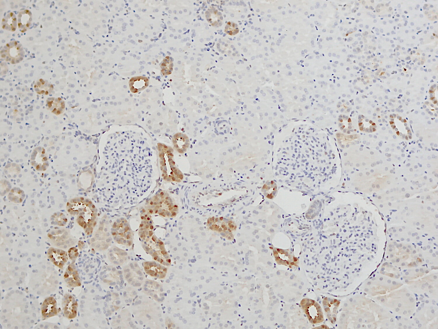
Galectin-3 (Gal-3, Mac-2) is a β-galactoside binding protein that is predominantly located in the cytoplasm and shuttles into the nucleus. In addition, it is secreted to the cell surface and into biological fluids. Depending on its localization, it exerts pleiotropic biological functions like cell growth, cell differentiation, inflammation, fibrogenesis, migration, apoptosis, and host defense (1). It is also involved in cancer pathogenesis, proliferation, and spreading of metastasis (2). Galectin-3 is highly expressed in the covering epithelia of the digestive tract and the urinary system (3) and plays important roles in the organization of renal and intestinal cells. Furthermore, Galectin-3 has been identified in a variety of other cell types including fibroblasts, keratinocytes, granulocytes, dendritic cells, and macrophages (2). Galectin-3 is expressed by certain tissue-resident macrophages such as Kupffer cells in the liver, red pulp macrophages in the spleen, and alveolar macrophages in the lung (3). In the brain, Galectin-3 is abundant in activated microglia but not expressed in resting cells. Following brain injury, Galectin-3 triggers anti-inflammatory properties of microglia (4). In Alzheimer’s disease, Gal-3 expression is strictly associated with microglial cell activation around Aβ plaques. In stroke, Gal-3 was found to be expressed primarily in proinflammatory microglial cells (5).
Certificates
ISO 9001 2015 Quality Management System and Green Lab Platinum certification level for sustaining laboratory processes.


Newsletter
Sign up for our newsletter and get the latest updates and news.