| Cat. No. HS-491 017 |
100 µg purified IgG, lyophilized. Albumin and azide were added for stabilization. For reconstitution add 100 µl H2O to get a 1mg/ml solution in PBS. Then aliquot and store at -20°C to -80°C until use. Antibodies should be stored at +4°C when still lyophilized. Do not freeze! |
| Applications | |
| Clone | SY-60F6 |
| Subtype | IgG2a (κ light chain) |
| Immunogen | Recombinant protein corresponding to residues near the central region of mouse FoxP3 (UniProt Id: Q99JB6) |
| Reactivity |
Reacts with: mouse (Q99JB6). Weaker signal: rat. No signal: human (Q9BZS1). Other species not tested yet. |
| Remarks |
IHC: Antigen retrieval with citrate buffer pH 6 is required. |
| Data sheet | hs-491_017.pdf |

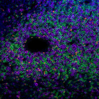
Detection of Foxp3+ CD4 regulatory T cells (red nucleus) in mouse spleen stained with anti-CD4 (green) and anti-CD8a (purple).
The transcription factor FoxP3 belongs to the family of forkhead transcription factors and controls the differentiation and function of regulatory T-cells (Tregs) (1). Tregs develop either in the thymus (tTreg), where they represent only ~2-3% of developing CD4+ thymocytes, or in the periphery by conversion of conventional CD4+ T cells into peripherally induced Treg (pTreg) cells (2). Expression of FoxP3 is induced by strong and persistent T cell receptor (TCR) signaling. FoxP3 binds DNA as homodimer or heterodimer with Foxp1 and interacts with other downstream transcription factors of TCR signaling, e.g., AP-1 transcription factors, NF-κB or Runx1 (1). Increasing evidence suggests that FoxP3 is expressed not only in Tregs, but also in a variety of tumor cells. However, tumor-FoxP3 has an inconsistent functional role and acts either as a tumor-suppressor or as a tumor-promoting factor (3). Mutations in FoxP3 lead to the severe autoimmune phenomena observed in patients with immune dysregulation, polyendocrinopathy, enteropathy, X-linked syndrome (IPEX) and in scurfy mice (4).