Cat. No.: HS-531 008
Amount: 50 µg
Price:
$420.00
| Cat. No. HS-531 008 |
50 µg purified recombinant IgG, lyophilized. Albumin and azide were added for stabilization. For reconstitution add 50 µl H2O to get a 1mg/ml solution in PBS. Then aliquot and store at -20°C to -80°C until use. Antibodies should be stored at +4°C when still lyophilized. Do not freeze! |
| Applications |
IP: not tested yet ICC: not tested yet IHC-P: 1 : 400 up to 1 : 1000 gallery ELISA: not tested yet |
| Clone | Rb-HNK-1 |
| Subtype | IgG1 (κ light chain) |
| Immunogen | Membrane extract of human lymphoblastoid cell line HSB-2 (UniProt Id: Q9P2W7) |
| Reactivity |
Reacts with: human (Q9P2W7). No signal: mouse, rat. Other species not tested yet. |
| Remarks |
This antibody is a chimeric antibody based on the monoclonal mouse antibody clone HNK-1. The constant regions of the heavy and light chains have been replaced with rabbit specific sequences. The antibody can therefore be used with standard anti-rabbit secondary reagents. The antibody has been expressed in mammalian cells. |
| Data sheet | Datasheet hs-531_008 |
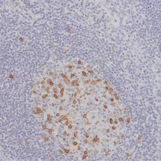
HNK-1 / CD57 positive cells are located in the germinal centers of the human tonsil
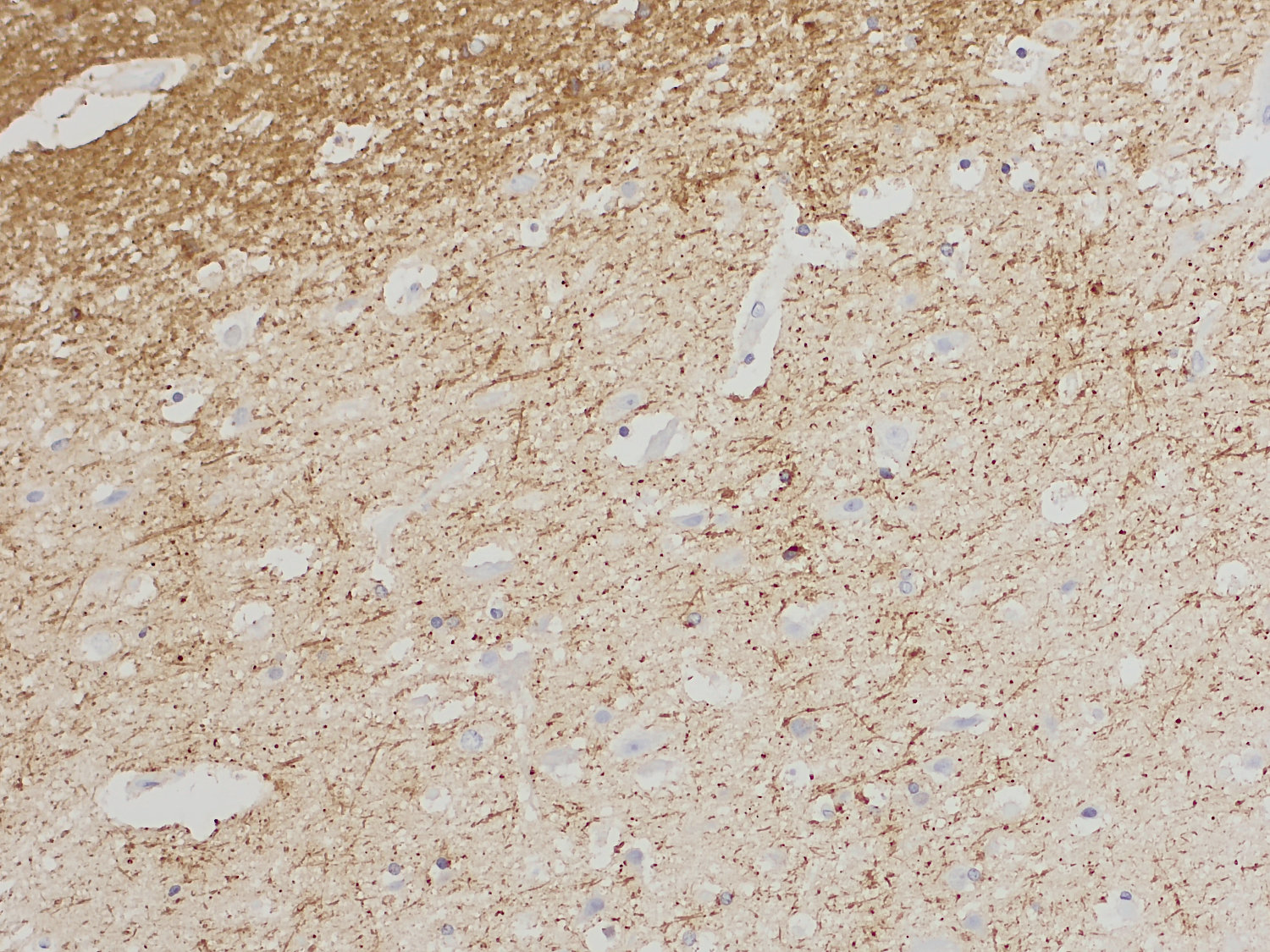
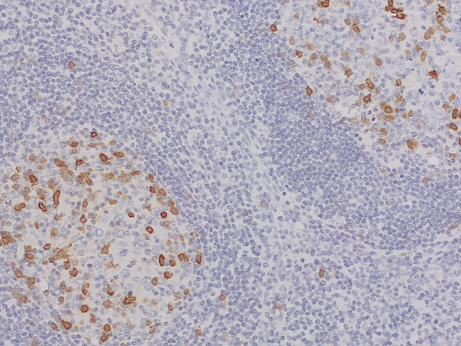
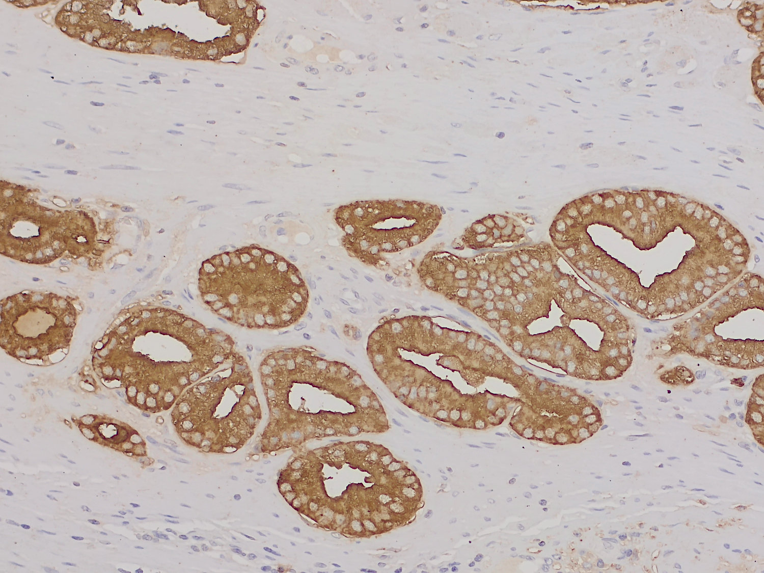
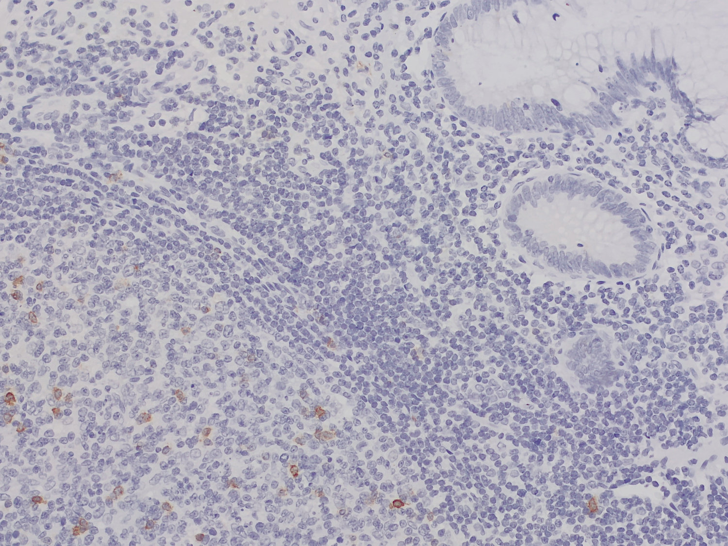
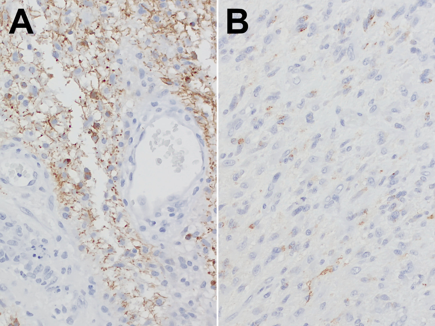
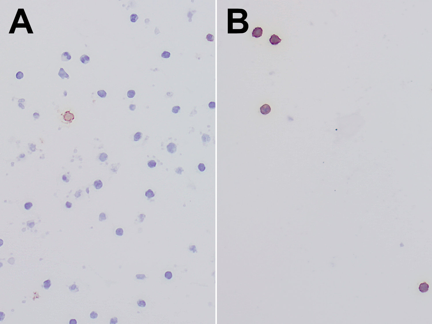
The human natural killer-1 (HNK-1) glyco-epitope, also known as LEU-7 or CD57, was first identified by a monoclonal antibody that recognizes a subset of human natural killer (NK) cells (1). This epitope features a unique trisaccharide structure, consisting of a 3-O-sulfated glucuronic acid linked to N-acetyl-lactosamine (HSO3-3GlcAß1-3Galß1-4GlcNAc-) and is primarily expressed on glycolipids and glycoproteins within the nervous system. Notable carriers are NCAM, L1, MAG, tenascin-R and tenascin-C (2). Studies in mice lacking HNK-1 expression reveal abnormal brain function, including impaired synaptic plasticity, deficits in dendritic spine maturation and disrupted spatial learning (2-4). In humans, HNK-1 is implicated in various central nervous system (CNS) disorders. For instance, autoantibodies targeting HNK-1 have been detected in certain neuropathies (5). Beyond its role in neural development, HNK-1 also appears to function as tumor suppressor. Elevated HNK-1 expression has been associated with improved survival outcomes in patients with prostate cancer or astrocytic tumors (6,7). In the field of immunology, HNK-1 is most commonly referred to as CD57. It is expressed on a subset of immune cells, particularly T cells and natural killer (NK) cells, and is associated with terminal differentiation and cellular senescence (8).
Certificates
ISO 9001 2015 Quality Management System and Green Lab Platinum certification level for sustaining laboratory processes.

Newsletter
Sign up for our newsletter and get the latest updates and news.