Cat. No.: HS-536 014
Amount: 100 µl
Price:
$355.00
| Cat. No. HS-536 014 |
100 µl antiserum, lyophilized. For reconstitution add 100 µl H2O, then aliquot and store at -20°C until use. Antibodies should be stored at +4°C when still lyophilized. Do not freeze! |
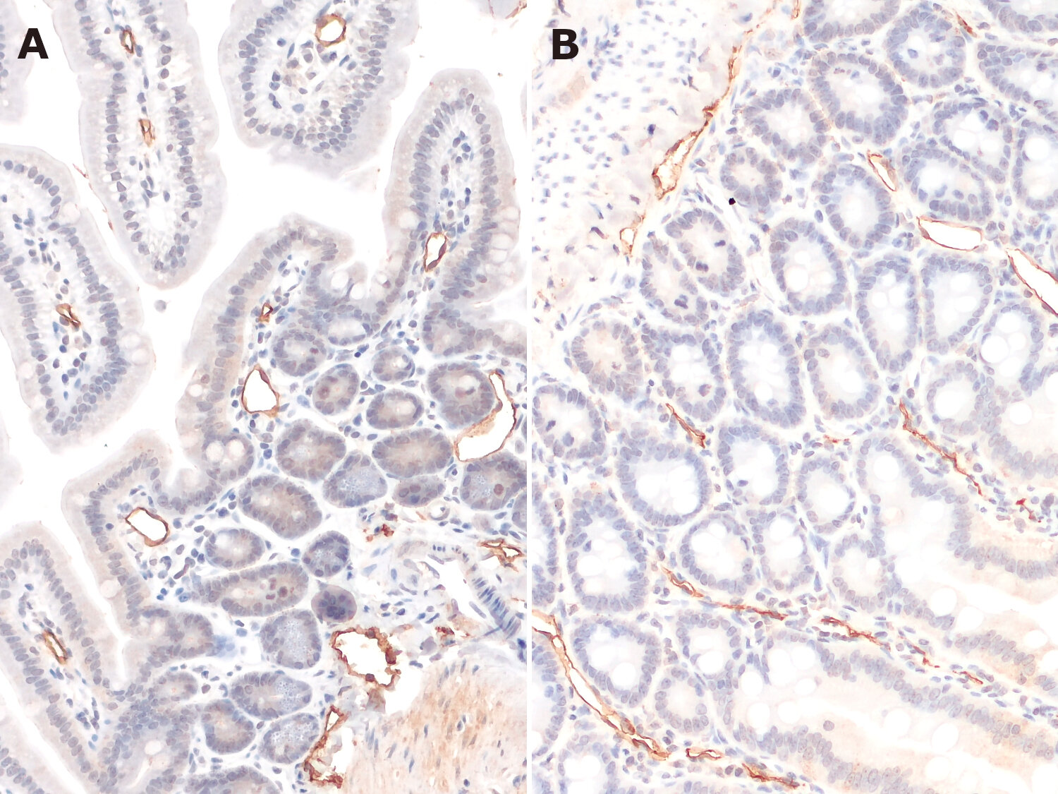
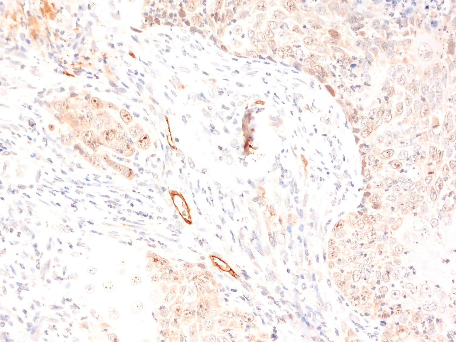
| Applications | |
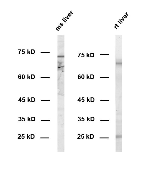
| Immunogen | Synthetic peptide corresponding to residues near the carboxy terminus of mouse Lyve1 (UniProt Id: Q8BHC0) |
| Reactivity |
Reacts with: mouse (Q8BHC0), rat (D3ZD19). No signal: human (Q9Y5Y7). Other species not tested yet. |
| Remarks |
IHC: Antigen retrieval with citrate buffer pH 6 can be applied to improve the signal to noise ratio. |
| Data sheet | Datasheet hs-536_014 |
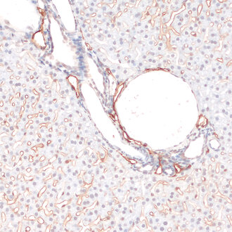
Transmembrane staining of Lyve1 in a mouse liver
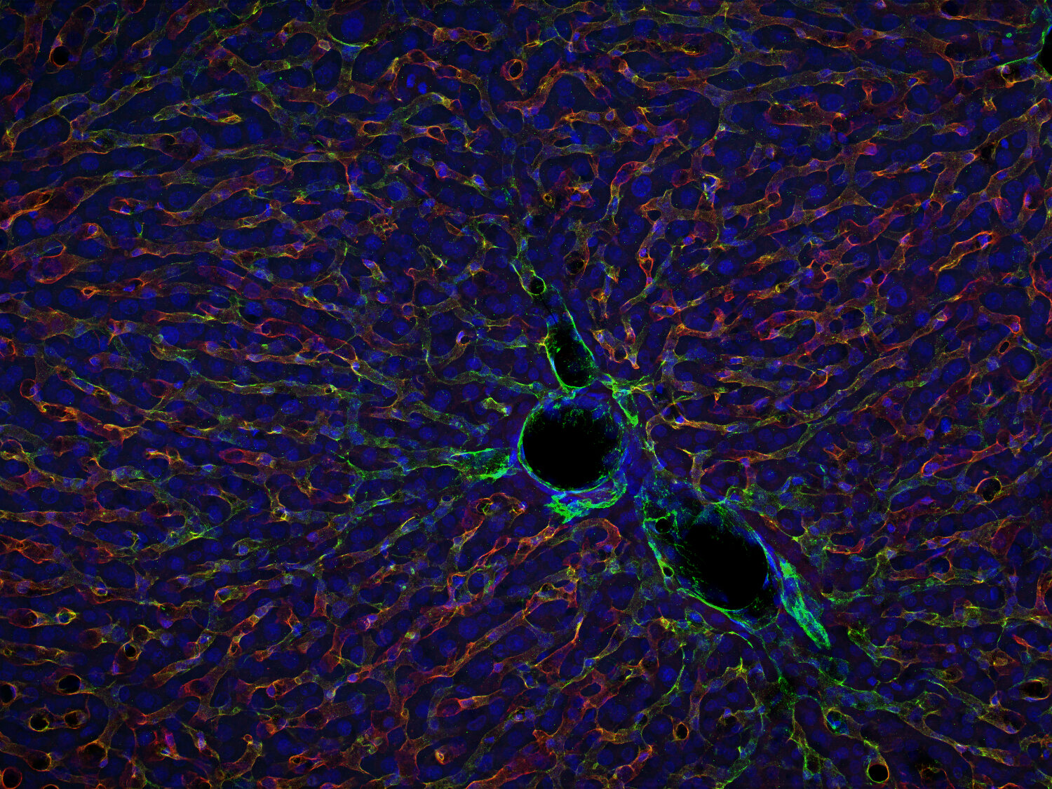
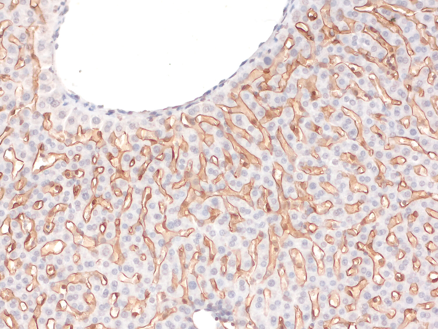
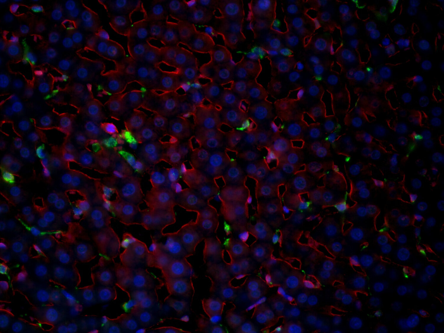
Lymphatic vessel endothelial hyaluronan receptor-1 (Lyve1) is a type I integral membrane glycoprotein and a member of the Link protein superfamily (1). It is predominantly expressed on lymphatic endothelial cells, where it functions as a key marker for identifying lymphatic vasculature and mediates binding and internalization of hyaluronan (HA) (2). Beyond the lymphatic system, Lyve1 is also detected in liver sinusoidal endothelial cells (3) and in subsets of tissue macrophages, including perivascular and interstitial macrophages in the lung, aorta, and mammary gland (4,5). Functionally, Lyve1 regulates HA metabolism, extracellular matrix remodeling, and leukocyte trafficking (2,4). Clinically, altered Lyve1 expression is implicated in several pathologies, as it correlates with lymphangiogenesis and metastatic spread in cancer (3,6), contributes to the regulation of vascular homeostasis and arterial stiffness (5), and participates in inflammatory and fibrotic conditions, such as rheumatoid arthritis and psoriasis (7).
Certificates
ISO 9001 2015 Quality Management System and Green Lab Platinum certification level for sustaining laboratory processes.
Newsletter
Sign up for our newsletter and get the latest updates and news.