HistoSure antibodies are specifically developed for use in formalin-fixed paraffin-embedded tissues and are extensively tested in a broad FFPE cell and tissue panel. Antibody testing in other applications (e.g. Western Blot, IHC, ICC) is an indispensable part of the validation process.
Specificity of antibodies are - if applicable - tested in formalin-fixed paraffin-embedded cell pellets of cells (over-) expressing the protein or protein fragment of interest. Whenever possible, the protein or proteinfragment is coupled to a reporter gene, to distinguish false positive from positive staining. Whenever possible, specificity of antibodies is also tested in cells or cell lines expressing the protein of interest at endogenous level.

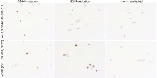
Figure 1: Anti-Histone H3.3 G34V (HS-388 011) staining in FFPE cell pellets transfected with H3.3 G34V-GFP construct (left column), H3.3 G34R-GFP construct (middle column) or non-transfected (right column) showing specificity for the G34V mutation.
Figure 1: Anti-Histone H3.3 G34V (HS-388 011) staining in FFPE cell pellets transfected with H3.3 G34V-GFP construct (left column), H3.3 G34R-GFP construct (middle column) or non-transfected (right column) showing specificity for the G34V mutation.
Tissue specificity and sensitivity of antibodies are tested using positive and negative tissues and whenever possible in high and low expressing tissues. For that purpose, SYSY has a comprehensive panel of mouse and rat tissues (e.g. brain, spleen, colon, thymus, heart, kidney, muscle, liver, pancreas…) and access to human clinical samples of various organs and indications. HistoSure antibodies display minimal staining artifacts and a high signal-to-noise ratio.


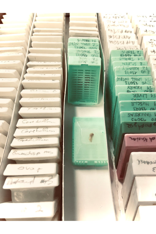
Figure 2: SYSY TissueBank of FFPE mouse tissues
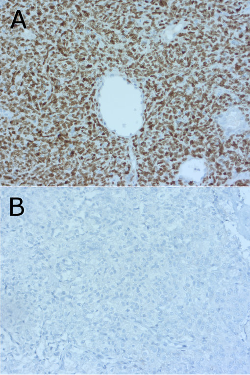
Figure 3: Detection of nuclear STAT6 (Cat. HS-378 011) in NAB2-STAT6 gene fusion positive (A, hemangiopericytoma) and negative (B, atypical meningioma) human FFPE tissues. Courtesy: Prof. Dr. F. Feuerhake and Prof. Dr. C. Hartmann, Dept. of Pathology, Neuropathology; Medizinische Hochschule Hannover