Immunodetection of Lyve1 in lymphatic and liver sinusoidal endothelial cells in a mouse liver.


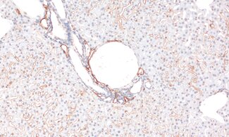
Immunodetection of Lyve1 in lymphatic and liver sinusoidal endothelial cells in a mouse liver.
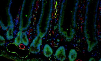
Immunohistochemical staining of a mouse small intestine section using Lyve1 (green) for lymphatic endothelial cells and CD31 (red) blood endothelial cells.
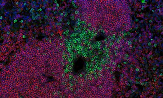
Immunohistochemical staining of a mouse spleen using anti-CD19 (red) and anti-CD3e (green) visualizes B-cell and T-cell populations.
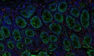
Immunohistochemical staining of a human small intestine section using anti-Galectin-3 (green) and anti-IBA1 (red) visualizes epithelial Galectin-3 and macrophages in the lamina propria.
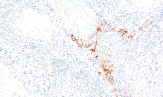
Detection of CD169-positive macrophages in a formalin fixed paraffin embedded (FFPE) human lymph node section.
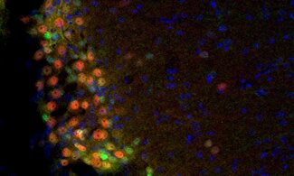
Immunohistochemical staining of a mouse olfactory bulb for T-bet (red) and Tbr2 (green) shows their expression in mitral and tufted cells.
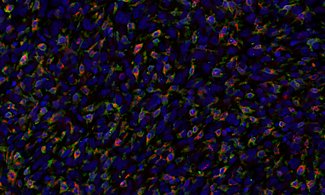
Immunohistochemical staining of a mouse syngeneic tumor using anti-CD68 (red) and anti-F4/80 (green) highlights infiltrating macrophages.
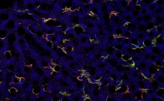
Immunodetection of CLEC4F-positive Kupffer cells (red) and F4/80-positive macrophages (green) in a mouse liver.
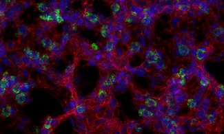
Immunohistochemical staining of a mouse lung section using anti-ICAM1 (red) and anti-LAMP3 (green) shows LAMP3-positive pneumocytes II within the alveolar epithelium.
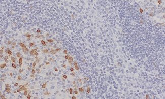
Indirect immunostaining of a human FFPE tonsil section shows that HNK-1 / CD57-positive cells are predominantly found in the germinal centers.
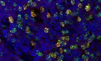
Immunohistochemical staining of a mouse spleen section using anti-Ly6G (red) and anti-CD11b (green) highlights Ly6G+ CD11b+ neutrophils.
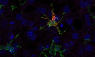
Immunohistochemical staining of mouse liver using anti-Chil3 (red) and anti-IBA1 (green) highlights M2 polarized Kupffer cells.

Immunodetection of Lyve1 in lymphatic and liver sinusoidal endothelial cells in a mouse liver.

Immunohistochemical staining of a mouse small intestine section using Lyve1 (green) for lymphatic endothelial cells and CD31 (red) blood endothelial cells.

Immunohistochemical staining of a mouse spleen using anti-CD19 (red) and anti-CD3e (green) visualizes B-cell and T-cell populations.

Immunohistochemical staining of a human small intestine section using anti-Galectin-3 (green) and anti-IBA1 (red) visualizes epithelial Galectin-3 and macrophages in the lamina propria.

Detection of CD169-positive macrophages in a formalin fixed paraffin embedded (FFPE) human lymph node section.

Immunohistochemical staining of a mouse olfactory bulb for T-bet (red) and Tbr2 (green) shows their expression in mitral and tufted cells.

Immunohistochemical staining of a mouse syngeneic tumor using anti-CD68 (red) and anti-F4/80 (green) highlights infiltrating macrophages.

Immunodetection of CLEC4F-positive Kupffer cells (red) and F4/80-positive macrophages (green) in a mouse liver.

Immunohistochemical staining of a mouse lung section using anti-ICAM1 (red) and anti-LAMP3 (green) shows LAMP3-positive pneumocytes II within the alveolar epithelium.

Indirect immunostaining of a human FFPE tonsil section shows that HNK-1 / CD57-positive cells are predominantly found in the germinal centers.

Immunohistochemical staining of a mouse spleen section using anti-Ly6G (red) and anti-CD11b (green) highlights Ly6G+ CD11b+ neutrophils.

Immunohistochemical staining of mouse liver using anti-Chil3 (red) and anti-IBA1 (green) highlights M2 polarized Kupffer cells.